equis - Articles
Skin: leukoderma


Introduction
- This is a common presentation in horses.
- Cause: it is secondary to many different conditions.
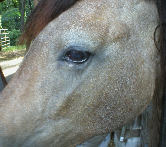
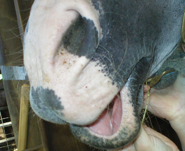
- Signs: leukoderma defines a depigmented area of the skin.
- Diagnosis: biopsy.
- Treatment: none.
- Prognosis: resolution depends on the underlying cause.
Presenting signs
- Patches of depigmented skin
 .
.
Breed/Species predisposition
- Gray horses are prone to developing depigmented areas.
Cost considerations
- Depends on underlying cause.
Pathogenesis
Etiology
- Multiple underlying causes can → leukoderma, including:
- Post-inflammatory depigmentation.
- Contact allergy Skin: contact dermatitis.
- Oncocherciasis Skin: onchocerciasis.
- Lupus erythematosus Systemic lupus erythematosus-like syndrome.
- Freezing and burns.
- Herpes coital exanthema Reproduction: coital exanthema - EHV 3.
- Pemphigus foliaceus Pemphigus foliaceus.
- Contact with rubber that contains phenols or monobenzyl ether of hydroquinone.
Pathophysiology
- Mechanism of depigmentation varies depending on the underlying cause.
- Melanocytes may be destroyed either by auto-immune mechanism or toxic.
- Also damages to the basement membrane → pigmentary incontinence (melanin drops in the dermis and is phagocytized by macrophages) which clinically → depigmentation.
Timecourse
- Depends on underlying cause.
Diagnosis
Subscribe To View
This article is available to subscribers.
Try a free trial today or contact us for more information.
Treatment
Subscribe To View
This article is available to subscribers.
Try a free trial today or contact us for more information.
Prevention
Subscribe To View
This article is available to subscribers.
Try a free trial today or contact us for more information.
Outcomes
Subscribe To View
This article is available to subscribers.
Try a free trial today or contact us for more information.
Further Reading
Publications
Refereed papers
- Recent references from PubMed and VetMedResource.
- Stannard AA (2000) Pigmentary disorders. Vet Derm 11 (3), 205-210 PubMed.
- Naughton G K, Mahaffey M & Bystryn J C (1986) Antibodies to surface antigens of pigmented cells in animals with vitiligo. Proc Soc Exp Biol Med 181 (3), 423-426 PubMed.
Other sources of information
- Scott D W & Miller W H (2011) Pigmentary Abnormalities. In: Equine Dermatology. 2nd edn. Saunders, USA. pp 389-397.
- Knottenbelt D C (2009) Ed Pascoe’s Principles and Practice of Equine Dermatology. 2nd edn. Saunders, USA.