canis - Articles
Peritoneal fluid: cytology


Overview
- Peritoneal fluid occurs within the abdominal/peritoneal cavity. Very little or no fluid can be aspirated unless effusion is present.
- Normal fluid is clear and colorless to slightly yellow, and is of low cellularity (<1000 nucleated cells/ml) and protein (<2.5 g/dl).
- Four mechanisms result in cavity effusions:
- Transudate - low specific gravity fluid crosses membrane barrier.
- Exudate - inflammation allows fluid with high cellular and protein component to cross vessel walls.
- Vessel or viscous rupture.
- Neoplastic proliferation.
- Cell characteristics vary in septic conditions and with neoplasia.
- Cytology enables finer differentiation of type of abdominal effusion Effusion: overview.
Uses
Alone
- Differentiate transudate from modified transudate or exudate.
- Differentiate septic from non-septic exudate.
- Diagnosis of lymphoma
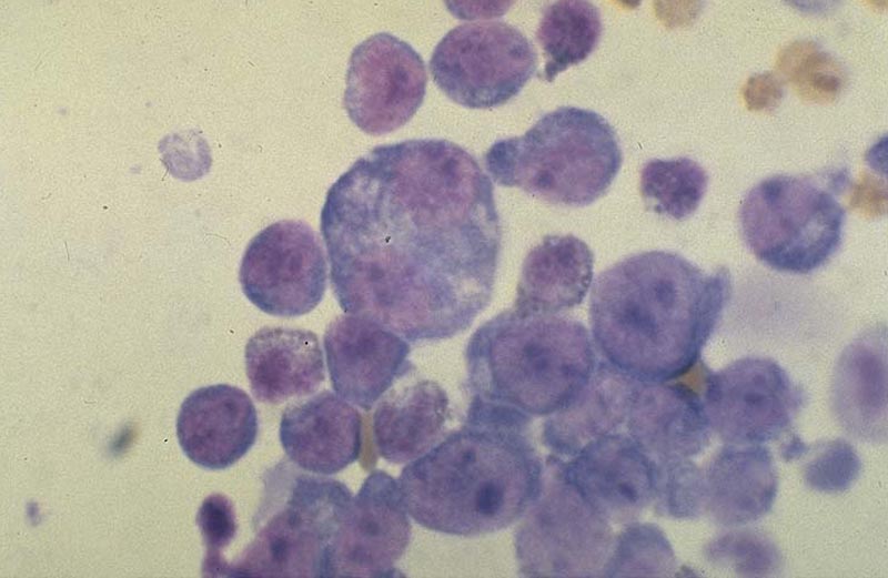 Lymphoma or mast cell tumor
 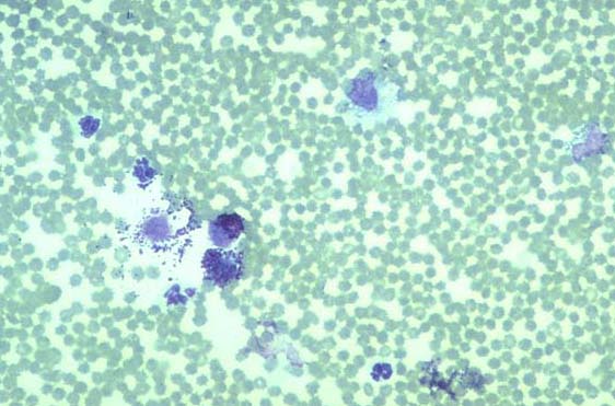 when neoplastic cells are exfoliated into the effusion.
In combination
- With other laboratory measures to diagnose cause of abdominal effusion, in particular to identify those due to infections or malignancy.
Other points
- Many tumors result in an effusion due to associated inflammation, eg with a necrotic tumor, or due to increased hydrostatic pressure but the tumor cells are not necessarilyexfoliated into the effusion.
Sampling
Subscribe To View
This article is available to subscribers.
Try a free trial today or contact us for more information.
Tests
Methodologies
- Centrifuge fluid at 1000 rpm for 10 min.
- Pour off supernatant and resuspend sediment by flicking tube.
- Put drop onto slide and make smear as for blood smear.
- Or use cytospin centrifuge.
- Air dry slide.
- Stain with Giemsa.
Availability
- Widely available at commercial laboratories.
- Smears can be examined in practice.
Validity
Sensitivity
- As with any cytologic specimen absence of evidence does not provide conclusive support for absence of a particular condition.
Specificity
- As with any cytologic specimen absence of evidence does not provide conclusive support for absence of a particular condition.
Technique intrinsic limitations
- Cytologic evaluation may be compromised by excessive blood contamination.
Technician extrinsic limitations
- May be difficult to differentiate reactive mesothelial cells from some neoplastic cells, eg carcinoma/adenocarcinoma/mesothelioma.
Result Data
Subscribe To View
This article is available to subscribers.
Try a free trial today or contact us for more information.
Further Reading
Publications
Refereed papers
- Recent references from VetMedResource and PubMed.
- Dunn J K & Villiers E (1998) Cytological and biochemical assessment of pleural and peritoneal effusions. In Pract 20, 501-505.
- Steyn P F & Wittum T E (1993) Radiographic epidiemiologic and clinical aspects of simultaneous pleural and peritoneal effusions in dogs and cats - 48 cases (1982-1991). JAAHA 202, 307-312.
- Wadle J R & Giger U (1990) Lipoprotein electrophoresis differentiation of chylous and nonchylous pleural effusions in dogs and cats and its correlation with pleural effusion triglycerole concentration. Comp Cont Ed Pract Vet 10, 121-136.
- Meyer D J & Franks P T (1987) Effusion - classification and cytologic examination. Comp Cont Ed Pract Vet 9, 123-128.
Other sources of information
- Cowell R L, Tyler R D & Meinkoth J H (1999) Diagnostic Cytology and Hematology of the Dog and Cat. 2nd edn. Mosby, USA.
- Duncan J R, Prasse K W & Mahaffey E A (1994) Veterinary Laboratory Medicine. Clinical Pathology. 3rd edn. Iowa University Press, USA.