canis - Articles
Dental trauma: tooth discoloration


Introduction
- Trauma commonly affects the oral cavity, this may be specific, eg dog catching a stone and fracturing a tooth or as part of a greater trauma syndrome, eg maxillofacial trauma from high rise or road traffic accidents.
- It is essential to fully assess the whole animal to ensure that life threatening aspects such as shock, hemorrhage or internal injury are identified and each insult accurately triaged.
- Dental, oral and maxillofacial trauma often exists in combination; if one injury is found there are frequently other injuries which may be less obvious.
- Examples include: tooth fracture (crown or root) Dental fracture Dental trauma: root fracture , tooth luxation Dental trauma: tooth luxation/avulsion , soft tissue laceration, jaw fracture, symphyseal separation, temporomandibular joint displacement Temporomandibular joint: luxation , oronasal communication, pulpal bleed (resulting in tooth discoloration), and pulpitis leading to pulpal necrosis also potentially causing tooth discoloration.
- Tooth discoloration: the result of a pulpal hemorrhage, pulpitis and pulpal necrosis secondary to tooth trauma. Other causes include hematogenous infection of the pulp, excessive orthodontic or occlusal forces or any event causing long term disruption to the blood supply to the tooth.
- Most commonly seen with canine teeth.
Presenting signs
- Tooth or teeth appear altered in color/ transparency.
- Color range: pink-purple, entire crown or crown tip, gray, beige-brown, reduced translucency.
- Crown color may develop and alter (usually darken) over weeks to months.
- Pain.
Acute presentation
- Oral/ dental discomfort/pain however signs are subtle and often missed.
- Tooth or teeth adjacent to an intact tooth (which is or is yet to discolor) may be fractured.
- Subluxation or soft tissue injury may be associated with the injured tooth.
- Tooth may regain normal color if pulpitis is reversible.
Age predisposition
- Traumatic injuries are usually considered more common in young dogs.
Cost considerations
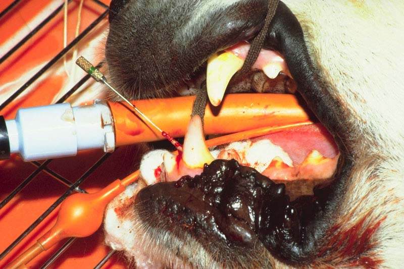
- Retention of affected teeth requires root canal therapy Endodontics: root canal therapy. A study has shown that the vast majority (about 94%) of discolored teeth have pulpal necrosis and that for a significant proportion of these there is no radiographic evidence of endodontic disease.
- Extraction Dental extraction of affected teeth may be the cheapest option but these teeth are usually immobile and present difficult extractions, thus this is not always the case.
- Monitor radiographs Radiology: dental do not give accurate information regarding toothache and require repeat procedures root canal therapy or extraction is thus generally preferred.
Special risks
- Ensure full assessment of all other injuries or insults
- Stabilize systemically first where required!
Pathogenesis
Etiology
- Blunt dental trauma.
Predisposing factors
General- Traumatic incident.
- Reduced alveolar support, eg pre-existing periodontitis Periodontitis.
Pathophysiology
- A blow to a tooth may cause trauma to the pulp, with resultant hemorrhage from pulpal vessels or pulpitis which may be reversible or if irreversible eventually progressing to pulpal necrosis
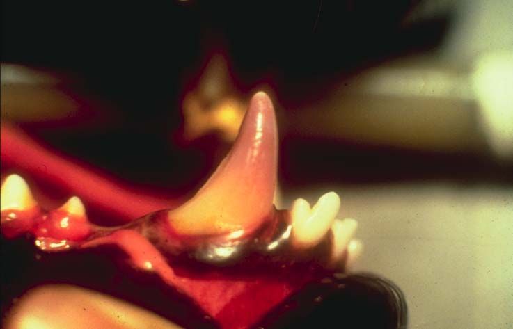
 . The blood hemoglobin breakdown products enter the dentinal tubules within the dentine of the crown and stain the dentine
 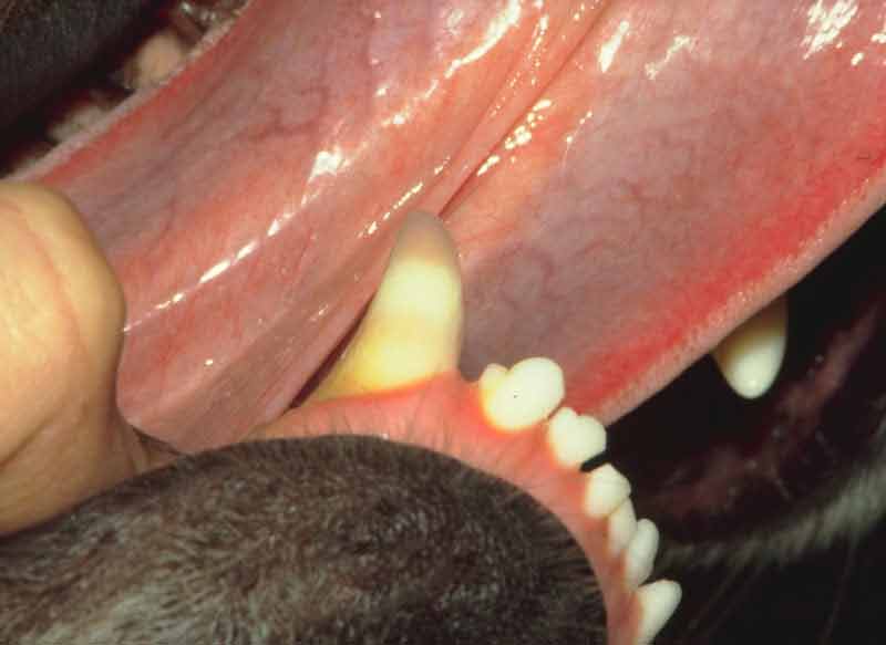 . This often begins as a pink discoloration several days after the trauma (this may return to normal if the initial discoloration is due to a reversible pulpitis), gradually changing to purple
 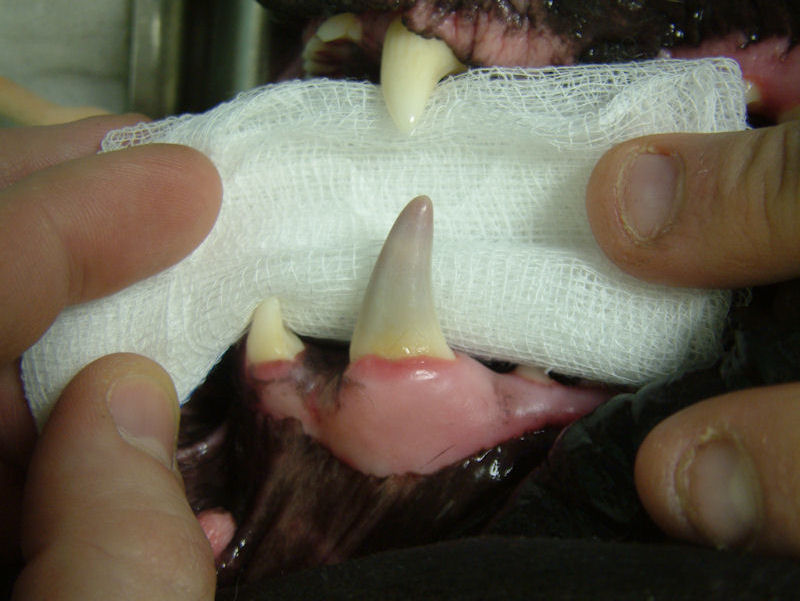 . A non-vital pulp will often lead to reduced translucency of the tooth, while a black necrotic pulp gives a gray appearance to the tooth. The majority of pulps become non-vital (94% reported as suffering irreversible pulpitis). Pulpitis produces toothache. Should non-vital pulps become infected, abscessation may occur. Bacteria colonizing a necrotic pulp may combine with iron from hemoglobin producing iron sulphide which may further darken the tooth.
Timecourse
- Acute incident however may not be noted for some time (many animals will continue to eat).
- Discoloration may alter over time, usually darkening as time from injury increases, but is usually permanent.
Diagnosis
Subscribe To View
This article is available to subscribers.
Try a free trial today or contact us for more information.
Treatment
Subscribe To View
This article is available to subscribers.
Try a free trial today or contact us for more information.
Prevention
Subscribe To View
This article is available to subscribers.
Try a free trial today or contact us for more information.
Outcomes
Subscribe To View
This article is available to subscribers.
Try a free trial today or contact us for more information.
Further Reading
Publications
Refereed papers
- Recent references from PubMed and VetMedResource.
- Hale F A (2001) Localized intrinsic staining of teeth due to pulpitis and pulp necrosis in dogs. J Vet Dent 18 (1), 14-20 PubMed.
Other sources of information
- Gorrel C (2004)Veterinary Dentistry for the General Practitioner.Saunders, Elsevier Science.
- Gorrel C, Penman S, Emily P (1993)Small Animal Oral Emergencies.Pergamon Press.
Organisation(s)
- The British Veterinary Dental Association:www.bvda.co.uk.