equis - Articles
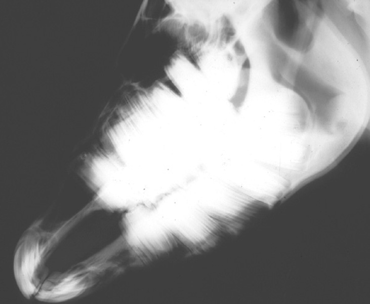
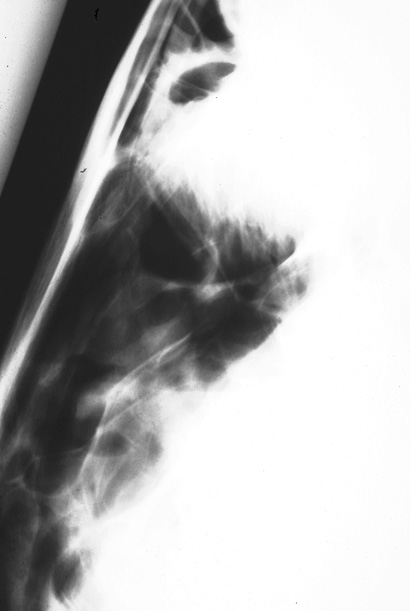
Head: radiography


Introduction
Radiography allows assessment of both osseous and soft tissue structures of the head Radiography: overview .
- Radiography of the skull is not difficult, however interpretation can be a problem as the anatomical structures are very complex.
Correct positioning will help considerably.
- See Head: radiology Head: radiology for interpretation.
Uses
- Diagnosis and evaluation of:
- Dental conditions, eg periodontal disease Teeth: periodontal disease , alveolar periostitis Mandible / maxilla: alveolar periostitis (chronic ossifying periostitis) , maleruption Teeth: maleruption , pseudocysts
 .
. - Fractures, eg mandible/maxilla Mandible / maxilla: fracture , skull fracture Head: fractures
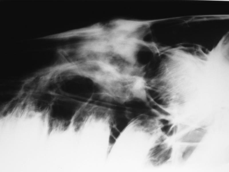 .
. - Paranasal sinus disease, eg bacterial sinusitis Paranasal sinuses: bacterial sinusitis , ethmoid hematoma Ethmoid: hematoma
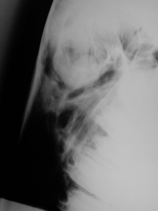 .
. - Guttural pouch disease, eg mycosis Guttural pouch: mycosis , bacterial or other infection Guttural pouch: empyema .
- Upper respiratory tract disorders, eg epiglottic entrapment Larynx: epiglottic entrapment , chondritis Larynx: arytenoid chondritis and calcification
 ; retropharyngeal infection, eg strangles Strangles (Streptococcus equi infection)
 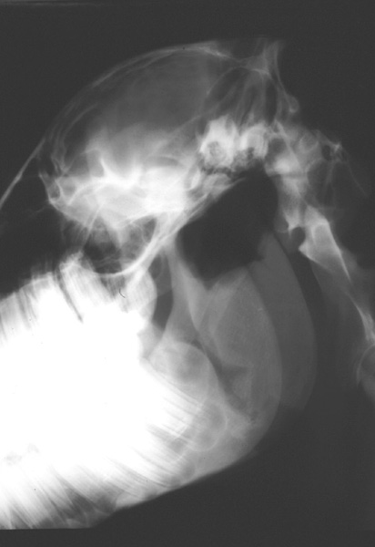 .
.
- Dental conditions, eg periodontal disease Teeth: periodontal disease , alveolar periostitis Mandible / maxilla: alveolar periostitis (chronic ossifying periostitis) , maleruption Teeth: maleruption , pseudocysts
Advantages
- Portable x-ray machines
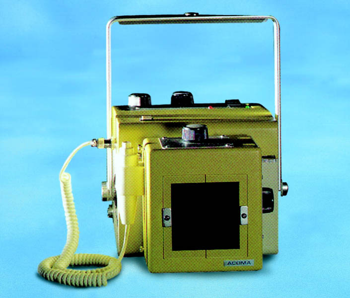 can be used for some views, particularly if fast rare-earth film screen combinations are used.
 - Dorsoventral views of the nasal septum can be obtained in a well-sedated standing horse Anesthesia: standing chemical restraint .
Disadvantages
- General anesthesia Anesthesia: general - overview is usually necessary for a good ventrodorsal view of the cranium.
Attention must be paid to radiation safety, use long handled or free standing cassette holders . Personnel holding the horse should wear lead gloves and aprons and use thyroid shields Radiography: radiation safety .
- Radiographic views and exposure techniques vary with the area of head being examined.
Exposure factors for the sinuses are considerably less than those for the teeth.
Alternative techniques
- Upper respiratory tract endoscopy Respiratory: endoscopy .
- Scintigraphy Bone: scintigraphy .
- Sinuscopy Paranasal sinus: sinuscopy .
- Exploratory surgery Respiratory: exploratory surgery .
Time required
Preparation
- 5-30 min depending on form of restraint.
Procedure
- 10-20 min.
Requirements
Subscribe To View
This article is available to subscribers.
Try a free trial today or contact us for more information.
Preparation
Subscribe To View
This article is available to subscribers.
Try a free trial today or contact us for more information.
Technique
Subscribe To View
This article is available to subscribers.
Try a free trial today or contact us for more information.
Aftercare
Subscribe To View
This article is available to subscribers.
Try a free trial today or contact us for more information.
Outcomes
Subscribe To View
This article is available to subscribers.
Try a free trial today or contact us for more information.
Further Reading
Publications
Refereed papers
- Recent references from PubMed and VetMedResource.
- Barakzai S (2014) Radiology of equine cheek teeth and sinus disorders. In Pract 36 (9), 466-472 BVA.
- Barrett M F & Easley J T (2013) Acquisition and interpretation of radiographs of the equine skull. Equine Vet Educ 25 (12), 643-652 VetMedResource.
- Barakzai S Z (2005) The equine paranasal sinuses Part 2: Radiography of the equine paranasal sinuses. UK Vet 10 (1), 5-9.
- Barakzai S Z & Weaver M P (2005) Imaging the equine temporohyoid region. Equine Vet Educ 17 (1), 14-15 VetMedResource.
- Barakzai S Z (2004) Equine dental radiography - Part 3: Radiographic signs of dental disease. UK Vet 9 (4), 14-17.
- Gibbs C (1996) Radiography of the head and soft tissues of the neck. Equine Vet Educ 3 VetMedResource.
- Park R D (1993) Radiographic examination of the equine head. Vet Clin North Am Equine Pract 9 (1), 49-74 PubMed.
Other sources of information
- Klugh D O (2003) Intraoral Radiography of Equine Premolars and Molars. In: Proc 49th AAEP Convention. pp 280-286.
- Butler J, Colles C M, Dyson S J, Kold S E & Poulos P W (1993) Clinical Radiology of the Horse. Blackwell, UK. ISBN 0-632-03271-5.